Hi, I used to use brainstorm to do locating ieeg. For MNE had this function with Python now, I tried it.
In brainstorm, when locating ieeg, we could enable “MIP: Functional”, then the electrodes are visable like this, which I think is better for locating.
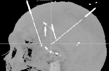
I’m not sure whether this is called a glass brain or what.
@alexrockhill may have some ideas here, I suppose!
Looks a bit like the bottom right plot in the GUI right now but with the CT. I’m guessing there is some automatic detection of the electrode contacts or marching cubes on the CT which can also be visualized but that is a direction for future development that I haven’t had time to work on enough yet.
If you select an electrode contact, you can see it on the 3D rendering of the GUI very similarly to this, I would suggest trying that, I rely heavily on that approach when I use the GUI to do localizations; without the 3D view it’s hard to really get your electrode contacts in a straight line and figure out which one is which.
Is there something that the image you attached allows you to do which the bottom right panel in this image does not?


And I think the way Brainstorm does it could have reference value.
For most of the electrodes, the length of space between each contact in the same shaft is static. So it would be nice if these parameters could be set

With these parameters, we could easily just choose the tip and the tail of the shaft, and then the coordinates of all the contacts would be calculated automatically.
Ah gotcha so the overall point is to have an automated process for selection. That is a future goal and I have worked on some approaches to doing that but it isn’t in the current version. I like your/the brainstorm idea of providing this interface but I think in practice, at least with the patients’ contacts I have localized, the spacing is not quite uniform (especially with the deepest and second deepest contacts) and there can be slight bending (or not so slight) of the electrode which would make me personally not want to use this feature especially when it only takes a few seconds to click roughly near the center of each one and use the snap-to-center feature (turned on). Not to mention, in our neurosurgical practice, electrodes with different contact sizes and spacings are used (which makes sense, you wouldn’t want to cover all areas with the same density).
I think that’s where this is headed to do as much as possible automatically, but the idea is to do it the best possible (as far current standards) way which requires validation and a lot of work so that will take some time.
Hi, may I ask what does snap-to-center function do?
It takes a nearby local maximum, finds all the voxels that decrease monotonicly and have intensities greater than half the max and then takes the centroid of those.